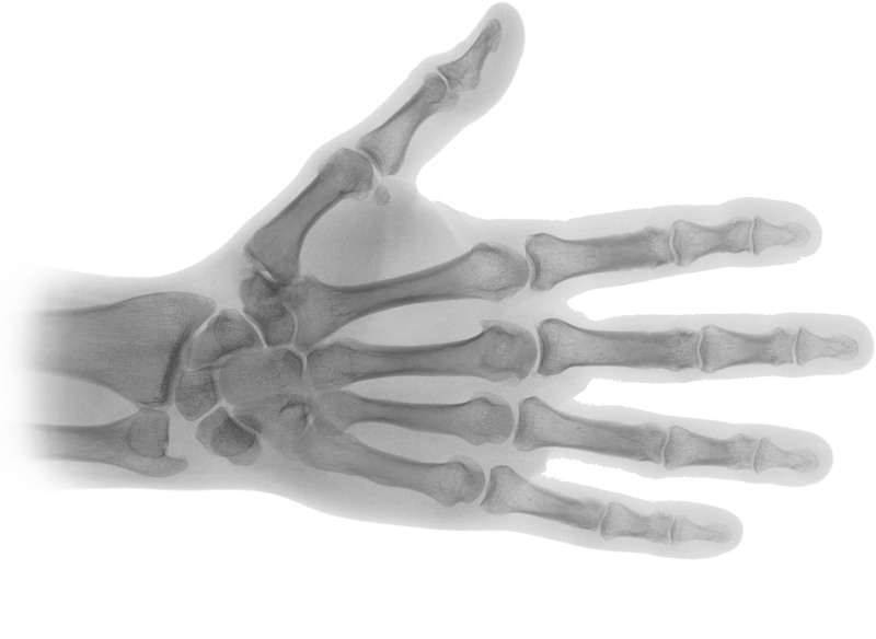
Download top and best high-quality free X-Ray PNG Transparent Images backgrounds available in various sizes. To view the full PNG size resolution click on any of the below image thumbnail.
License Info: Creative Commons 4.0 BY-NC
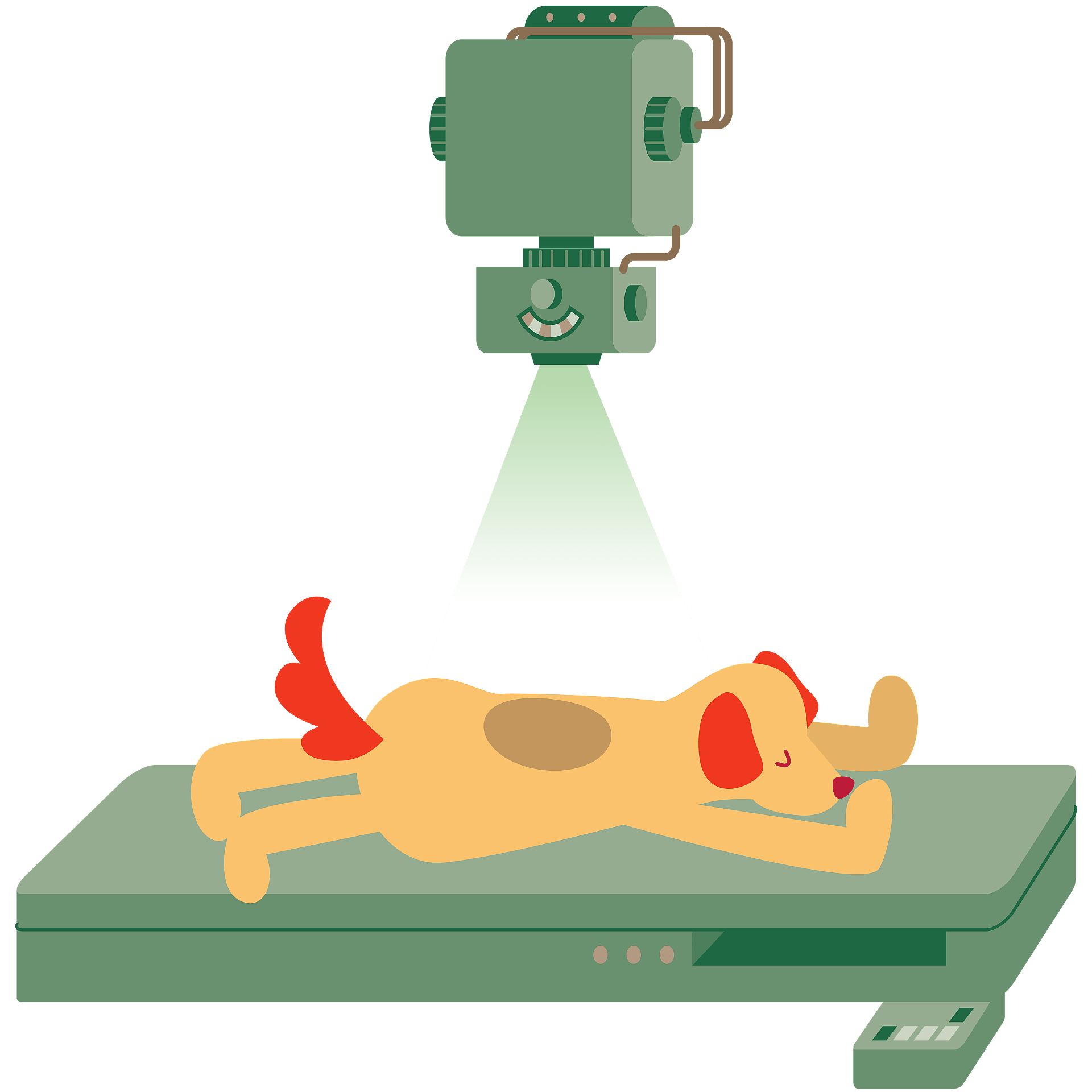
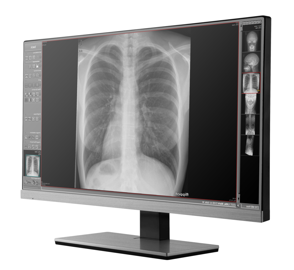
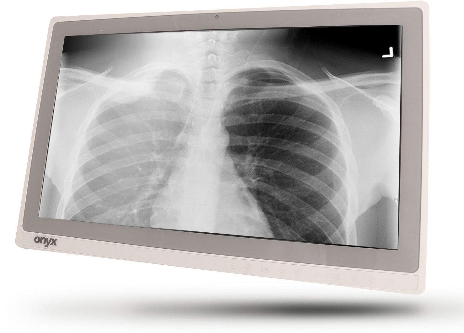
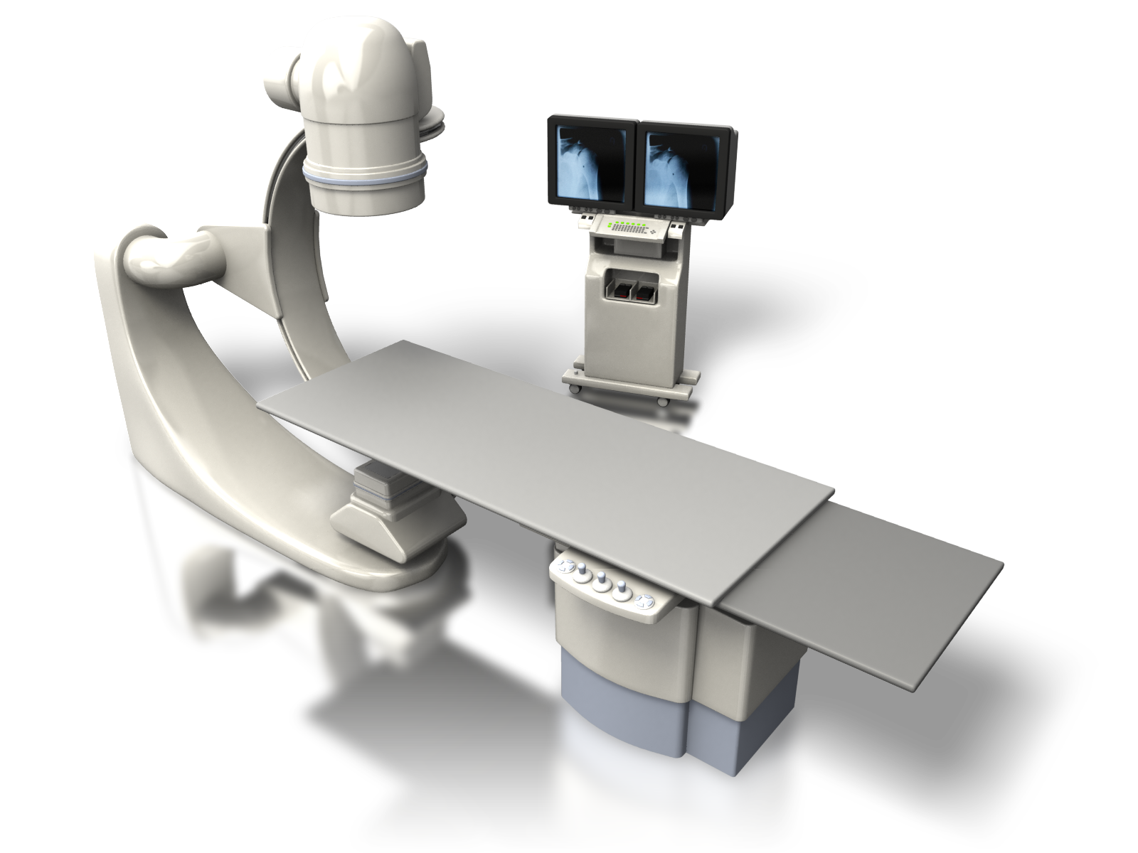
X-rays make up X-radiation, a form of high energy electromagnetic radiation. Most X-rays have a wavelength ranging from 0.1 to 10 nanometers, corresponding to frequencies between 30 petahertz and 30 exahertz (3×10¹⁶ Hz to 3×10¹⁹ Hz) and energies between 100 eV and 200 keV. The wavelengths of X-rays are shorter than those of UV rays and generally longer than those of gamma rays. In many languages, X-ray radiation is called Röntgen radiation, after the German scientist Wilhelm Röntgen, who discovered it on November 8, 1895. He called it X-ray to signify an unknown type of radiation. The spelling of X-rays in English includes the variants x-ray(s), xray(s), and X ray(s).
Before their discovery in 1895, X-rays were only one type of unidentified radiation emanating from experimental discharge tubes. They were noticed by scientists investigating the cathode rays produced by these tubes, which are beams of energetic electrons which were observed for the first time in 1869. Many of the first Crookes tubes (invented around 1875) have without any doubt radiated from X-rays because early researchers noticed effects that were attributable to them, as detailed below. The Crookes tubes created free electrons by ionization of the residual air in the tube by a high DC voltage of anywhere between a few kilovolts and 100 kV. This voltage accelerated the electrons from the cathode to a speed high enough to create X-rays when they hit the anode or the glass wall of the tube.
The first experimenter to have (without knowing it) produced X-rays was the actuary William Morgan. In 1785, he presented an paper to the Royal Society of London describing the effects of the passage of electric currents through a partially evacuated glass tube, producing a glow created by X-rays. This work was further explored by Humphry Davy and his assistant Michael Faraday.
When Stanford University, a professor of physics at Stanford University, created his “electric photography”, he also generated and detected X-rays without knowledge. From 1886 to 1888, he studied at the Hermann Helmholtz laboratory in Berlin, where he became familiar with the cathode rays generated in vacuum tubes when a voltage was applied through separate electrodes, as previously studied by Heinrich Hertz and Philipp Lenard. His letter of January 6, 1893 (describing his discovery as “electric photography”) to The Physical Review was duly published and an article entitled Without Lens or Light, Photographs Taken with Plate and Object in the Dark appeared in the San Francisco Examiner.
Starting in 1888, Philipp Lenard conducted experiments to see if cathode rays could pass from the Crookes tube into the air. He built a Crookes tube with a thin aluminum “window” at the end, facing the cathode so that the cathode rays hit it (later called a “Lenard tube”). He discovered that something had happened that would expose the photographic plates and cause fluorescence. He measured the power of penetration of these rays through different materials. It has been suggested that at least some of these “Lenard rays” are actually X-rays.
Download X-Ray PNG images transparent gallery.
- X Ray
Resolution: 1280 × 721
Size: 63 KB
Image Format: .png
Download
- X Ray PNG Clipart
Resolution: 2000 × 2000
Size: 233 KB
Image Format: .png
Download
- X Ray PNG Download Image
Resolution: 980 × 980
Size: 72 KB
Image Format: .png
Download
- X Ray PNG File Download Free
Resolution: 943 × 1149
Size: 849 KB
Image Format: .png
Download
- X Ray PNG File
Resolution: 1920 × 1920
Size: 40 KB
Image Format: .png
Download
- X Ray PNG Free Download
Resolution: 2000 × 2857
Size: 38 KB
Image Format: .png
Download
- X Ray PNG Free Image
Resolution: 696 × 1500
Size: 291 KB
Image Format: .png
Download
- X Ray PNG HD Image
Resolution: 1000 × 916
Size: 350 KB
Image Format: .png
Download
- X Ray PNG High Quality Image
Resolution: 604 × 396
Size: 244 KB
Image Format: .png
Download
- X Ray PNG Image File
Resolution: 968 × 433
Size: 204 KB
Image Format: .png
Download
- X Ray PNG Image HD
Resolution: 600 × 543
Size: 31 KB
Image Format: .png
Download
- X Ray PNG Image
Resolution: 1479 × 1062
Size: 1292 KB
Image Format: .png
Download
- X Ray PNG Images
Resolution: 1600 × 1200
Size: 859 KB
Image Format: .png
Download
- X Ray PNG Photo
Resolution: 1200 × 1200
Size: 702 KB
Image Format: .png
Download
- X Ray PNG Pic
Resolution: 981 × 800
Size: 42 KB
Image Format: .png
Download
- X Ray PNG Picture
Resolution: 840 × 747
Size: 104 KB
Image Format: .png
Download
- X Ray PNG Transparent HD Photo
Resolution: 800 × 584
Size: 195 KB
Image Format: .png
Download
- X Ray PNG
Resolution: 596 × 680
Size: 452 KB
Image Format: .png
Download
- X Ray Transparent
Resolution: 512 × 512
Size: 21 KB
Image Format: .png
Download

