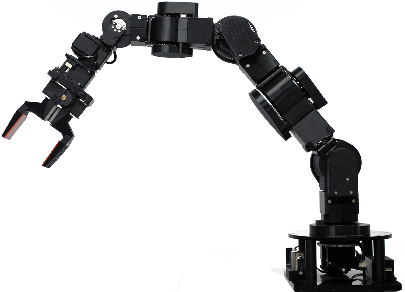
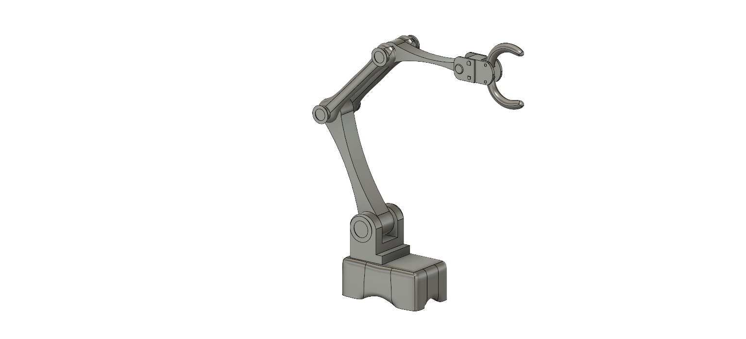
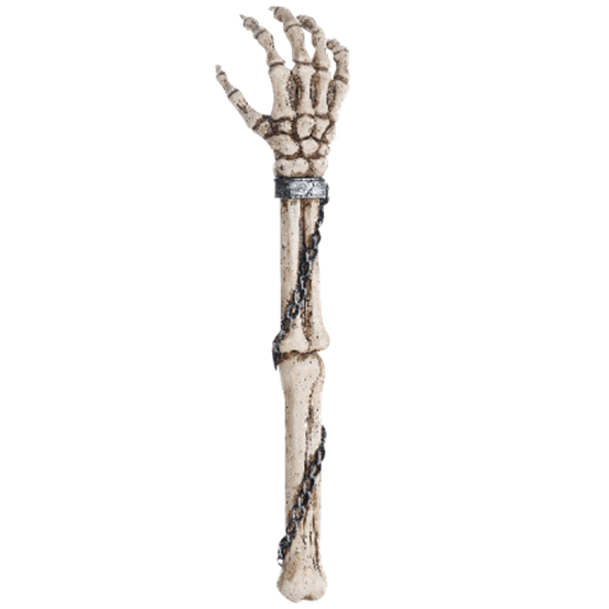
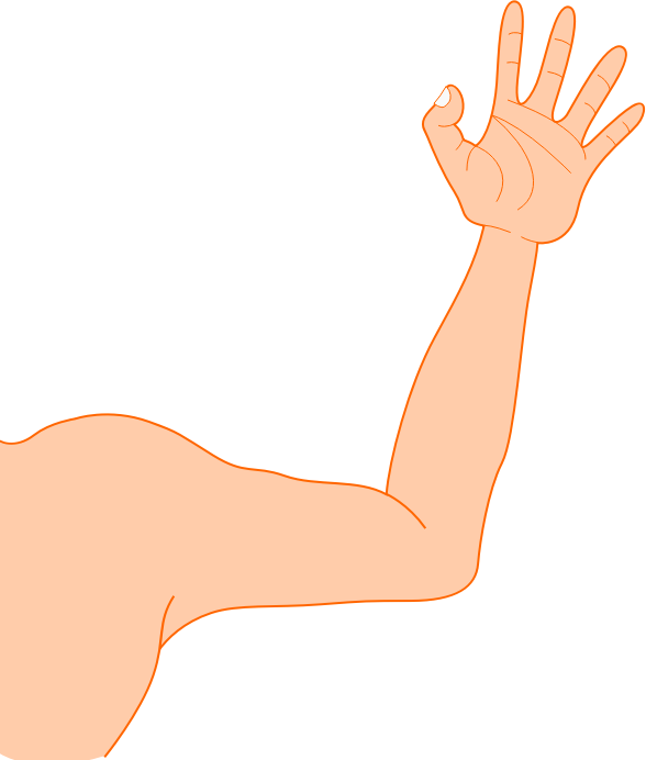
Download top and best high-quality free Arm PNG Transparent Images backgrounds available in various sizes. To view the full PNG size resolution click on any of the below image thumbnail.
License Info: Creative Commons 4.0 BY-NC
The arm is the portion of the upper limb between the glenohumeral joint and the elbow joint in human anatomy. The arm is commonly extended through the hand. It is split into three sections:
- The upper arm (from the shoulder to the elbow)
- The forearm (from the elbow to the hand)
- The hand
The shoulder girdle, with its bones and muscles, is anatomically defined a component of the arm. The Latin word brachium can refer to either the entire arm or just the upper arm.
One of the three long bones of the arm is the humerus. At the shoulder joint, it connects with the scapula, and at the elbow joint, it connects with the two long bones of the arm, the ulna and radius. The elbow is a complicated hinge joint that connects the humerus’s end to the radius and ulna’s ends.
The arm is divided into two osteofascial compartments by a fascial layer (lateral and medial intermuscular septa) that separates the muscles into two osteofascial compartments: the anterior and posterior compartments. The periosteum (outer bone layer) of the humerus combines with the fascia.
Three muscles make up the anterior compartment: the biceps brachii, brachialis, and coracobrachialis. The musculocutaneous nerve innervates them all. The triceps brachii muscle supplied by the radial nerve is the solitary muscle in the posterior compartment.
The musculocutaneous nerve, which runs from C5 to C7, is the major supply source for anterior compartment muscles. It comes from the brachial plexus’s lateral cord of nerves. It pierces the coracobrachialis muscle and sends branches to the brachialis and biceps brachii muscles. The anterior cutaneous nerve of the forearm is where it ends.
The radial nerve prolongs the posterior cord of the brachial plexus that runs from the fifth cervical spinal nerve to the first thoracic spinal nerve. This nerve penetrates the lower triangle area of the arm. It lies deep to the triceps brachii (an imagined space limited by, among others, the shaft of the humerus and the triceps brachii). It passes via the radial groove of the humerus with the deep artery of the arm. This is significant clinically because a fracture of the bone shaft here might induce lesions or even nerve transections.
The brachial artery is the major artery in the arm. This artery branches out from the axillary artery. The transition from axillary to brachial occurs distal to the lower boundary of the teres major. The deep artery of the arm is a minor branch of the brachial artery. This branching occurs immediately below teres major’s bottom boundary.
The brachial artery travels through the anterior compartment of the arm to the cubital fossa. Like the median nerve and basilic vein, it runs on a plane between the biceps and triceps muscles. Venae comitantes accompanies it (accompanying veins). It provides branches to the anterior compartment muscles. In the cubital fossa, the artery runs between the median nerve and the biceps muscle tendon. The pain then spreads to the forearm.
The radial nerve and the deep artery of the arm pass via the lower triangular region. It has a close association with the radial nerve from here on out. They are both positioned on the spiral groove of the humerus, deep within the triceps muscle. As a result, a bone fracture can cause radial nerve damage and haematoma in the arm’s internal tissues. The artery subsequently anastamoses with the brachial artery’s recurrent radial branch, giving a diffuse blood supply for the elbow joint.
Download Arm PNG images transparent gallery.
- Arm Background PNG
Resolution: 512 × 512
Size: 8 KB
Image Format: .png
Download
- Arm No Background
Resolution: 766 × 662
Size: 504 KB
Image Format: .png
Download
- Arm PNG Clipart
Resolution: 2055 × 2400
Size: 354 KB
Image Format: .png
Download
- Arm PNG Cutout
Resolution: 1686 × 2244
Size: 104 KB
Image Format: .png
Download
- Arm PNG File
Resolution: 805 × 581
Size: 113 KB
Image Format: .png
Download
- Arm PNG Free Image
Resolution: 1649 × 1722
Size: 1777 KB
Image Format: .png
Download
- Arm PNG HD Image
Resolution: 480 × 596
Size: 30 KB
Image Format: .png
Download
- Arm PNG Image File
Resolution: 1536 × 698
Size: 97 KB
Image Format: .png
Download
- Arm PNG Image HD
Resolution: 800 × 420
Size: 28 KB
Image Format: .png
Download
- Arm PNG Image
Resolution: 960 × 574
Size: 80 KB
Image Format: .png
Download
- Arm PNG Images HD
Resolution: 561 × 685
Size: 233 KB
Image Format: .png
Download
- Arm PNG Images
Resolution: 800 × 800
Size: 67 KB
Image Format: .png
Download
- Arm PNG Photo
Resolution: 1111 × 1280
Size: 124 KB
Image Format: .png
Download
- Arm PNG Photos
Resolution: 512 × 512
Size: 21 KB
Image Format: .png
Download
- Arm PNG Pic
Resolution: 555 × 555
Size: 78 KB
Image Format: .png
Download
- Arm PNG Picture
Resolution: 588 × 692
Size: 29 KB
Image Format: .png
Download
- Arm PNG
Resolution: 2663 × 2789
Size: 231 KB
Image Format: .png
Download
- Arm Transparent
Resolution: 625 × 720
Size: 46 KB
Image Format: .png
Download
- Arm
Resolution: 1664 × 1929
Size: 88 KB
Image Format: .png
Download


