Download top and best high-quality free Anatomy Heart PNG Transparent Images backgrounds available in various sizes. To view the full PNG size resolution click on any of the below image thumbnail.
License Info: Creative Commons 4.0 BY-NC
When we think of the heart, we often imagine a symbol of love and emotion. However, the heart is more than just a metaphor. It is a vital organ that pumps blood and provides oxygen and nutrients to all parts of our body. In this article, we will explore the anatomy of the heart and its functions.
The Structure of the Heart
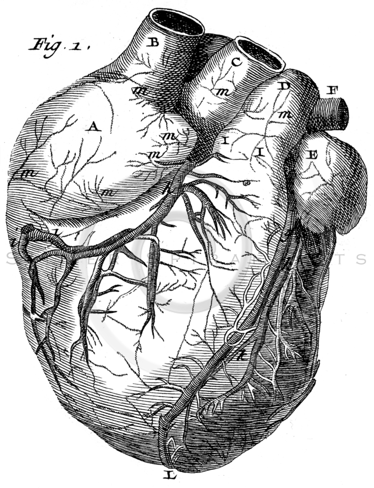
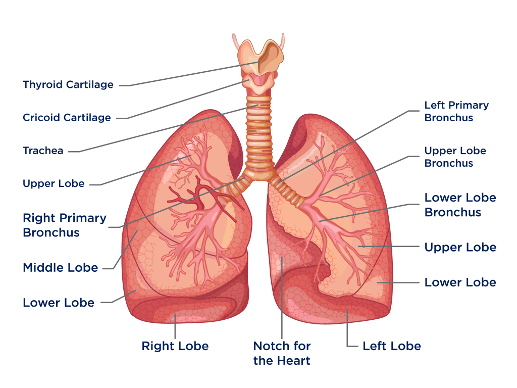
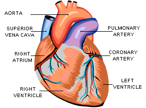
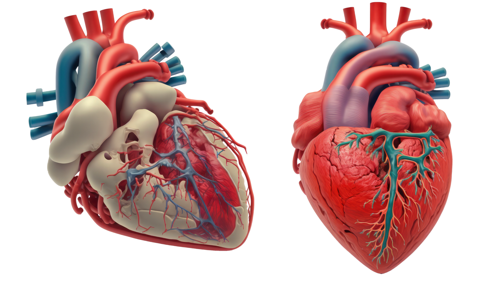
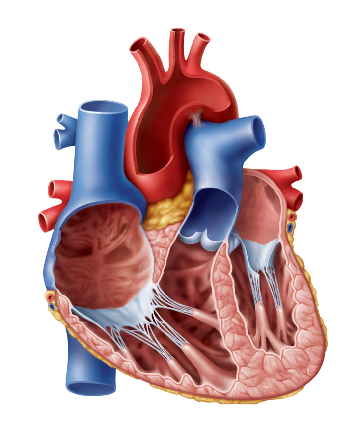
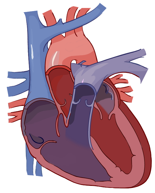
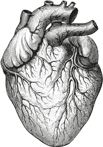
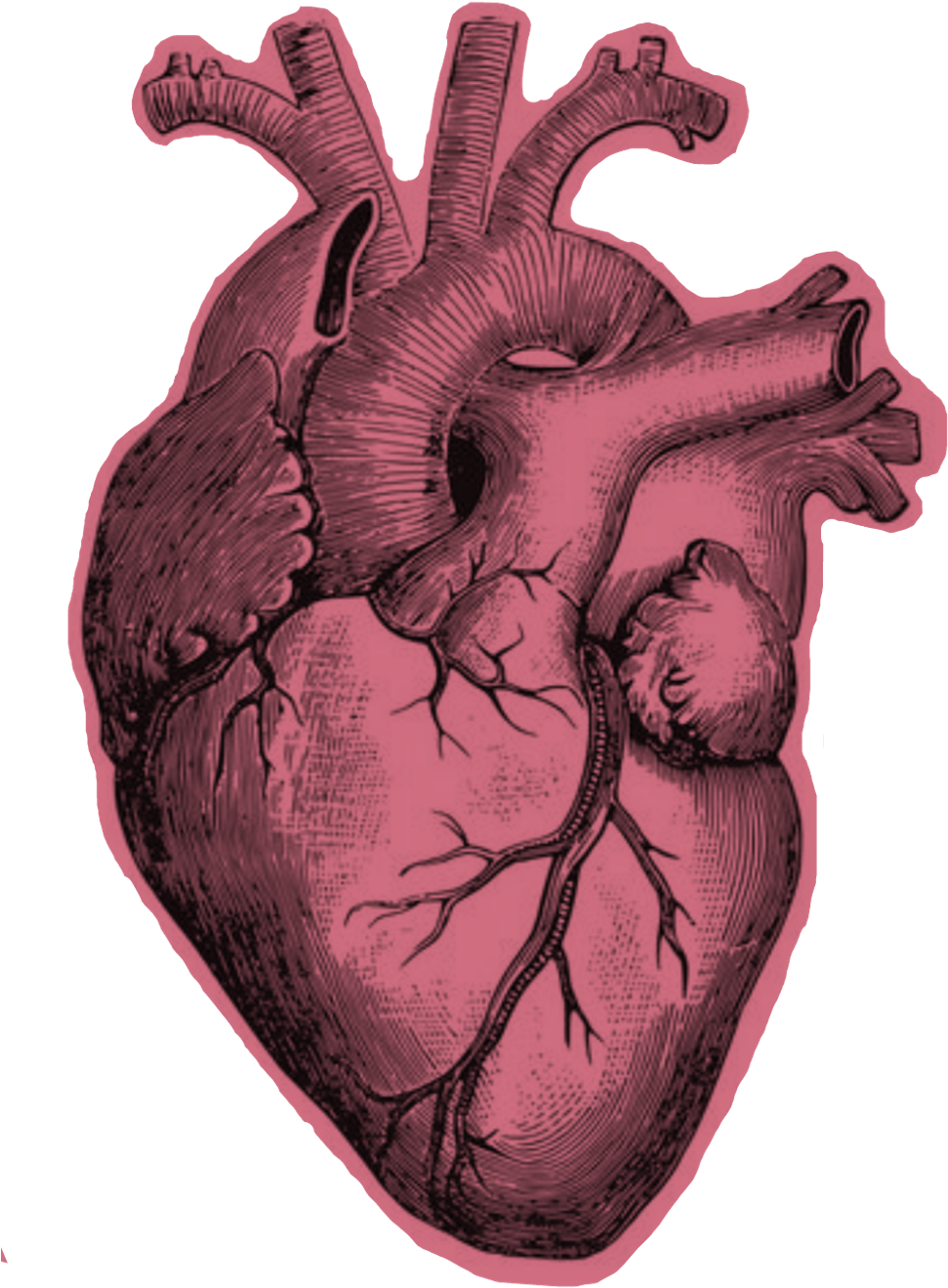
The heart is a muscular organ that is approximately the size of a human fist. It is located in the chest, between the lungs, and is protected by the ribcage. The heart is divided into four chambers, including the right atrium and ventricle, and the left atrium and ventricle. These chambers are separated by valves, which ensure that the blood flows in the right direction.
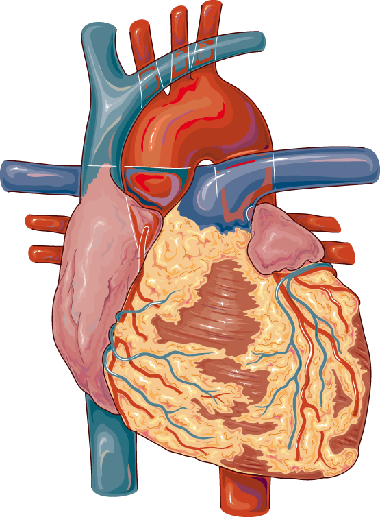
The heart is also surrounded by the pericardium, a sac-like membrane that contains fluid to lubricate and protect the heart. The pericardium has two layers, the parietal layer, which lines the fibrous sac, and the visceral layer, which covers the heart.
The Functions of the Heart
The primary function of the heart is to pump blood throughout the body. The blood carries oxygen and nutrients to the tissues and organs and removes waste products, such as carbon dioxide. The heart has a complex electrical system that controls the rate and rhythm of the heartbeat. The sinoatrial (SA) node, located in the right atrium, is known as the pacemaker of the heart. It generates electrical impulses that spread across the atria and stimulate the contraction of the atrial muscles. The impulses then travel to the atrioventricular (AV) node, which delays the impulse for a brief period to allow the atria to empty completely before the ventricles contract. The impulse then passes to the Bundle of His, a collection of specialized heart cells that conduct the electrical signal to the Purkinje fibers, causing the ventricles to contract.
The Blood Vessels of the Heart
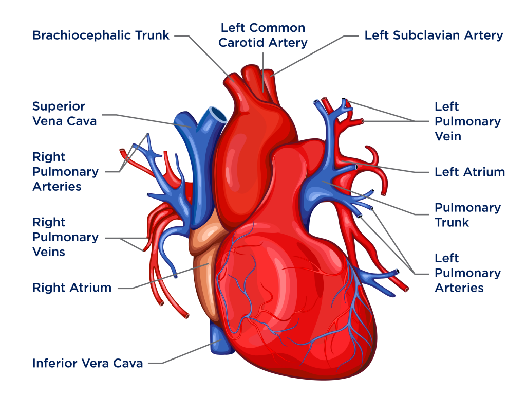
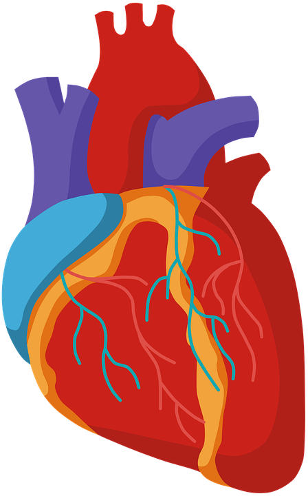
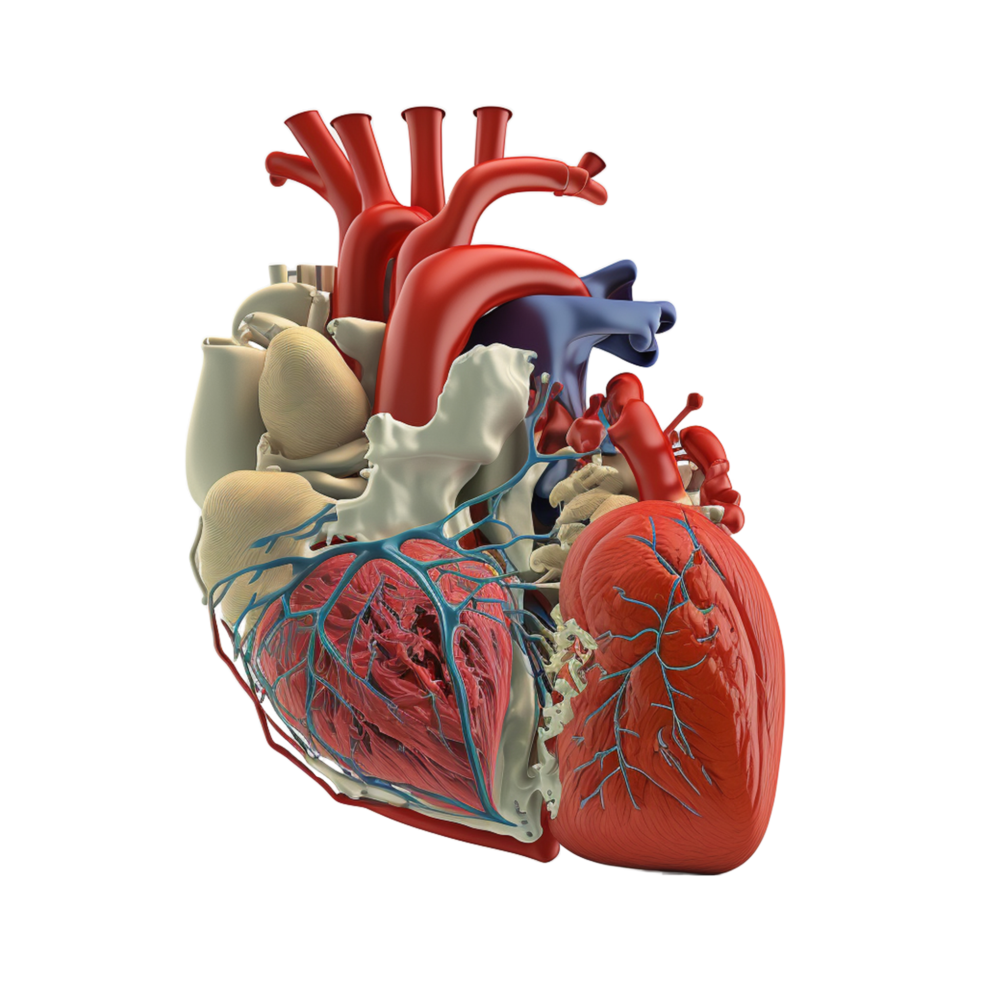
The heart is supplied with blood by a network of blood vessels, including the coronary arteries and veins. The coronary arteries arise from the aorta and branch out to supply blood to the heart muscle. The coronary veins drain the blood from the heart muscle and return it to the right atrium. Blockage or narrowing of these vessels can lead to coronary artery disease, which can result in a heart attack.
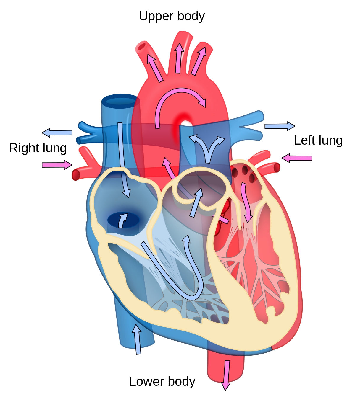
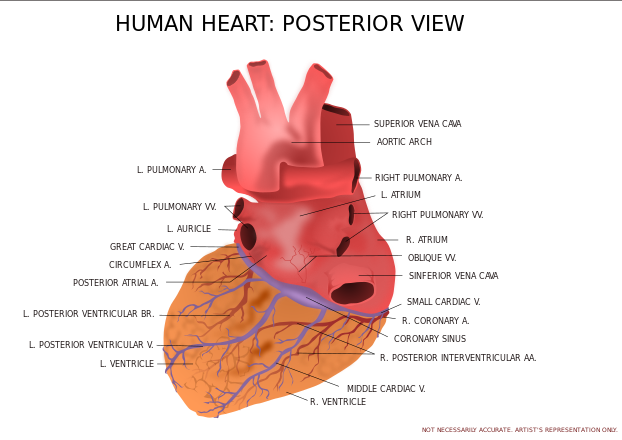
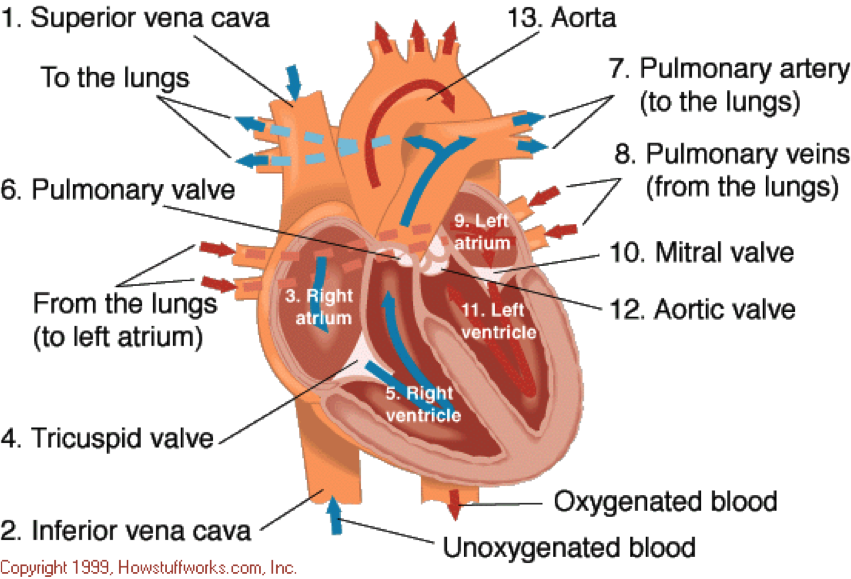
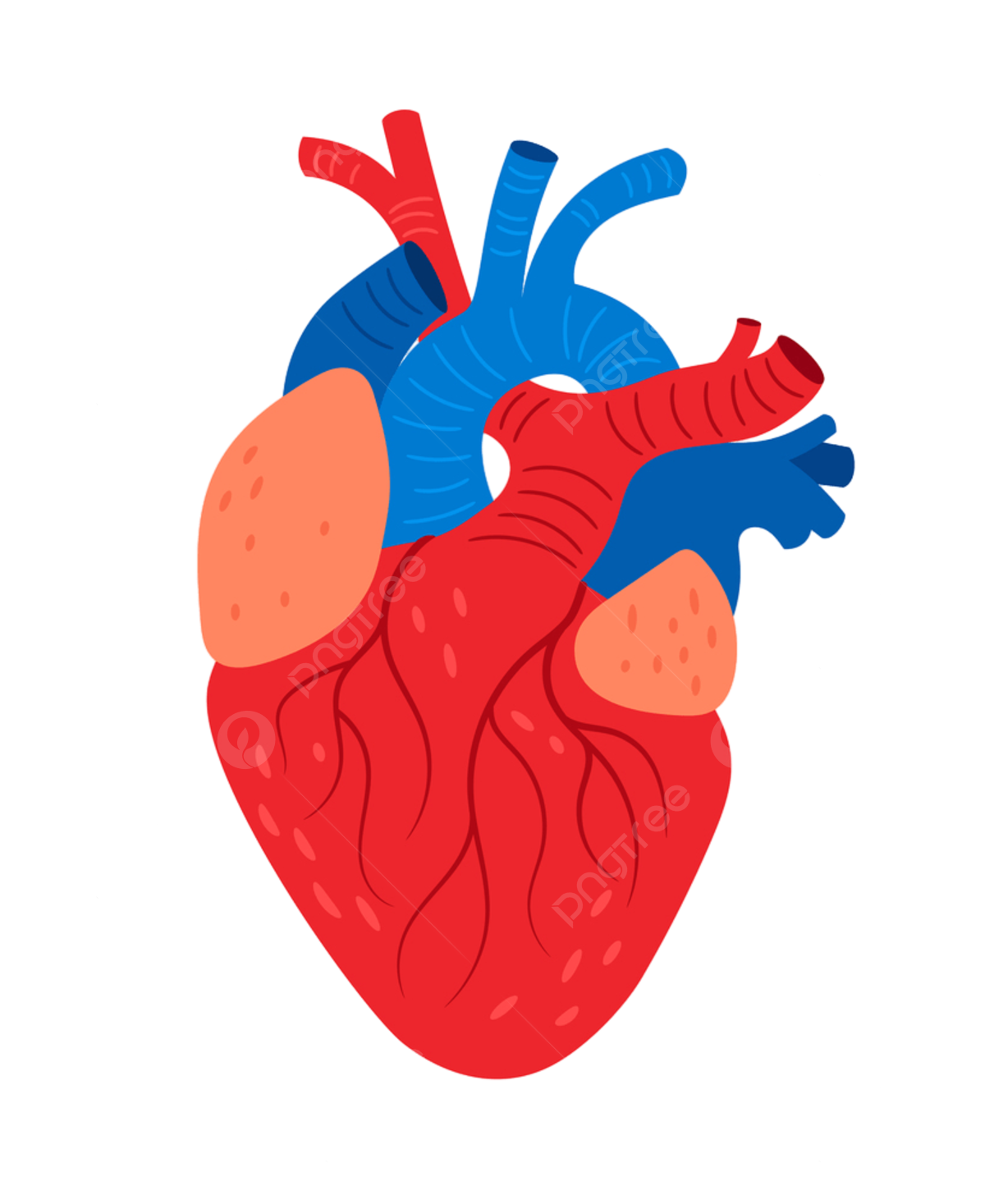
The heart also receives blood from the systemic circulation. The superior and inferior vena cava bring deoxygenated blood from the body to the right atrium. The blood passes through the tricuspid valve into the right ventricle, which pumps it through the pulmonary valve and into the pulmonary artery. The pulmonary artery carries the blood to the lungs, where it is oxygenated. The oxygenated blood then returns to the heart through the pulmonary veins and enters the left atrium. The blood then flows through the mitral valve into the left ventricle, which pumps it through the aortic valve and into the aorta. The aorta branches out to supply oxygenated blood to the organs and tissues of the body.
The heart is a remarkable organ that is critical to the functioning of our entire body. Understanding its structure, functions, and blood vessels can give us insight into how it works and what we can do to maintain its health. By taking care of our heart through a healthy diet, regular exercise, and medical attention when necessary, we can ensure that it will continue to beat strong for years to come.
Download Anatomy Heart PNG images transparent gallery
-

- Anatomy Heart PNG Picture
Resolution: 736 × 960
Size: 523 KB
Image Format: .png
Download
-

- Anatomy Heart PNG
Resolution: 1024 × 768
Size: 326 KB
Image Format: .png
Download
-
- Anatomy Heart Transparent
Resolution: 1024 × 768
Size: 295 KB
Image Format: .png
Download
-

- Anatomy Heart

Resolution: 896 × 1280
Size: 315 KB
Image Format: .png
Download
-

- Anatomy Heart Background PNG
Resolution: 555 × 754
Size: 265 KB
Image Format: .png
Download
-

- Anatomy Heart No Background
Resolution: 293 × 218
Size: 17 KB
Image Format: .png
Download
-

- Anatomy Heart PNG Background
Resolution: 1200 × 1345
Size: 518 KB
Image Format: .png
Download
-

- Anatomy Heart PNG Clipart
Resolution: 448 × 720
Size: 126 KB
Image Format: .png
Download
-

- Anatomy Heart PNG Cutout
Resolution: 980 × 980
Size: 680 KB
Image Format: .png
Download
-

- Anatomy Heart PNG File
Resolution: 1698 × 980
Size: 1398 KB
Image Format: .png
Download
-

- Anatomy Heart PNG Free Image
Resolution: 1280 × 1339
Size: 2847 KB
Image Format: .png
Download
-

- Anatomy Heart PNG HD Image
Resolution: 539 × 800
Size: 265 KB
Image Format: .png
Download
-

- Anatomy Heart PNG Image File
Resolution: 622 × 440
Size: 111 KB
Image Format: .png
Download
-

- Anatomy Heart PNG Image HD
Resolution: 729 × 874
Size: 716 KB
Image Format: .png
Download
-

- Anatomy Heart PNG Image
Resolution: 851 × 579
Size: 361 KB
Image Format: .png
Download
-

- Anatomy Heart PNG Images HD
Resolution: 1920 × 1920
Size: 2273 KB
Image Format: .png
Download
-

- Anatomy Heart PNG Images
Resolution: 546 × 678
Size: 122 KB
Image Format: .png
Download
-

- Anatomy Heart PNG Photo
Resolution: 418 × 597
Size: 231 KB
Image Format: .png
Download
-

- Anatomy Heart PNG Photos
Resolution: 1200 × 1467
Size: 443 KB
Image Format: .png
Download
-

- Anatomy Heart PNG Pic
Resolution: 971 × 1320
Size: 1288 KB
Image Format: .png
Download